Gold dyes can be green
The vibrant color of turmeric has made it useful since ancient times as a cooking spice and clothing dye. There has been much interest in researching it’s potential as a disease therapy. This research focus has prompted labs to apply this low cost, non-toxic substance to tissue staining (1) and bioimaging (2) studies. While turmeric has not been certified by the Biological Stain Commission just yet, studies have demonstrated some good potential to replace synthetic dyes.
Histology methods require lab safety and economizing
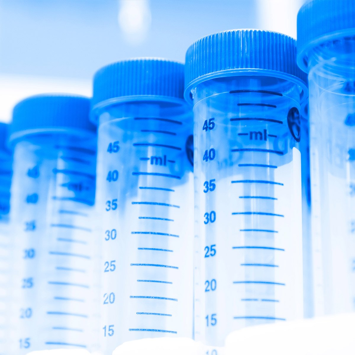
Histochemical methods are used by thousands of labs in both medicine and in scientific research. There is a certain amount of artistry involved to these techniques. There are hundreds of methods to fix, embed and stain tissue sections. Good slide results depend on understanding of the potential effects to tisues from each step in processing.
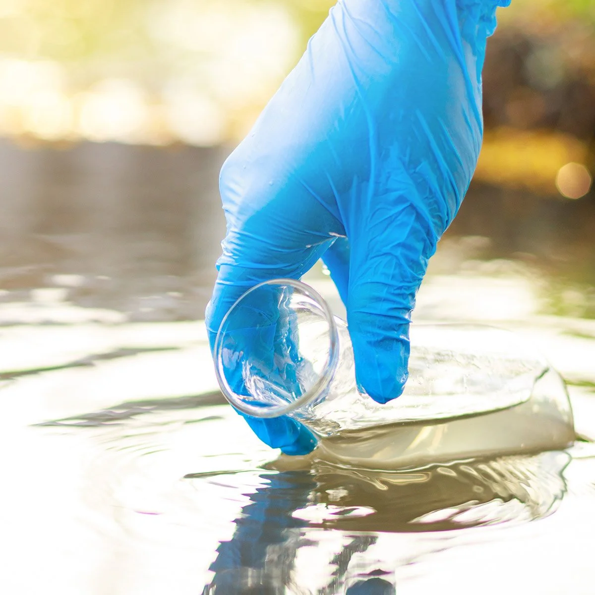
Toxicity is a commonly accepted hazard in histological processing. Xylene and toluene solvents have neurotoxic effects. Formaldehyde fixative is a known carcinogen Recently, there has been interest in reducing the occupational hazards of histology lab work by replacing chemical hazards with bio-friendly alternatives (3). In some cases, natural compounds also provide the bonus of being less costly. This work is a natural fit to improve lab sustainability.
H&E staining
The most popular histology staining method worldwide is “H&E”. Hematoxylin and eosin stains are considered a diagnostic gold standard for pathologists. Hematoxylin solutions contain haematein dye and metal ions that produce blue staining of cell nuclei. These solutions more so stain the cell chromatin and are not specific to DNA itself. The dye is derived from the central american logwood tree, then oxidized with a chemical oxidant. Eosin is a highly pH dependent synthetic dye, derived from fluorescein, that counterstains tissue sections pink. A quality Eosin stain should show at least three shades from light pink to bright fuschia Most of the time paraffin wax embedded tissue samples are used, but sometimes H&E is used on frozen tissue sections.
What do scientists know so far about turmeric’s unique staining features?
The primary compound in turmeric, curcumin, has some unique features. Curcumin regulates the action of cellular membrane proteins indirectly by changing the physical properties of membranes. You might think of it as molecular thickener. As a medical therapy, turmeric’s limitation has been that it is not bioavailable, because it is not water soluble. Just the opposite, curcumin is lipophilic. It could possibly be classified as a lysochrome. A lysochrome is not really a dye, but rather a colored, non-polar substance that is dissolves in lipids, (i.e. the commonly used Sudan Black B stain).
Although eosin is the most efficient counterstain for hematoxylin, turmeric can also be effectively used as an alternative for eosin (1). In sharp contrast to eosin, curcumin lacks pH sensitivity. That’s certainly a practical feature. Curcumin is non-toxic and eco-friendly so disposal is easy. It would be interesting to see turmeric stained frozen section tested by histologists. Frozen sections are are commonly used by histology and pathology labs when lipid component preservation is required - or when results are needed fast. Hopefully it would work great! Could it be used for high volume throughput paraffin sections? It appears to dye uniformly, so its a possibility.
Curcumin also fluoresces. Most synthetic fluorescent dyes are carcinogenic and expensive. Their toxicity makes it tricky to get an accurate image of the active processes in living cells because they die immediately after the application. Scientists at Quanzhou Normal University have used nanotechnology to compensate for turmeric’s low stability and water solubility. By encapsulation with biodegradable PLGA nanoparticles, curcumin was demonstrated to be an excellent fluorescent probe for bioimaging living cells.
“Unlike conventional chromophores such as organic dyes and quantum dots which are limited by photo bleaching and toxicity, natural available curcumin is strongly fluorescent and nontoxic, making it suitable for bio-imaging applications. “ - Minghuan Liu et al.
Will turmeric staining catch on? It fits a major principal of laboratory safety, which is to substitute less toxic materials whenever possible. We hope that more optimized methods will be established. Until then, please continue to protect your health in histology labs and prevent any ecological impacts by not pouring histochemicals down drains. Thank you for being “labconscious”!
Read more:
Hema Suryawanshi et al. Curcuma longa extract – Haldi: A safe, eco-friendly natural cytoplasmic stain (2017)J Oral Maxillofac Pathol.
Minghuan Liu et al. Polymeric Encapsulation of Turmeric Extract for Bioimaging and Antimicrobial Applications (2018) Macromolecular Rapid Communications
NaAnanthalakshmi Ramamoorthy et al. Natural Alternatives for Chemicals Used in Histopathology Lab- A Literature Review (2016) J Clin Diagn Res.